
Note: This article is for educational purposes only. It explains heart pictures, diagrams, and basic anatomy in plain American English. It is not a substitute for medical advice, diagnosis, or treatment from a qualified healthcare professional.
The human heart is often drawn as a cute red symbol on greeting cards, but the real thing is far more impressive. It is a muscular, four-chambered pump that works every second of your life, even when you are asleep, daydreaming, or pretending not to hear your alarm. Heart pictures and diagrams help turn this busy organ from a mysterious “thump-thump machine” into something you can actually understand.
Whether you are studying biology, reading a medical report, preparing a health article, or simply trying to understand what your doctor is pointing at on a screen, learning heart anatomy is incredibly useful. A good heart diagram shows the chambers, valves, major blood vessels, coronary arteries, and blood-flow direction. Once those pieces click together, the heart stops looking like a complicated plumbing project and starts making beautiful sense.
What Is the Heart?
The heart is a hollow muscular organ located in the chest, slightly left of center. Its main job is to pump blood through the circulatory system. Blood carries oxygen and nutrients to the body’s tissues and helps carry away waste products. In simple terms, the heart is the body’s delivery driver, pickup service, traffic controller, and power pump all in one compact package.
In most adults, the heart is roughly the size of a closed fist. But do not let the size fool you. This fist-sized organ keeps blood moving through thousands of miles of blood vessels. It sends blood to the lungs to pick up oxygen, then pumps oxygen-rich blood to the brain, muscles, organs, skin, and every other tissue that likes staying alivewhich is, thankfully, all of them.
Why Heart Pictures and Diagrams Matter
Heart pictures make anatomy easier to understand because they show relationships. Reading “the right ventricle pumps blood to the pulmonary artery” is helpful, but seeing it on a labeled heart diagram is much better. Visuals help you understand where the chambers sit, how valves guide blood, and why the left side of the heart is built for high-pressure pumping.
There are several common types of heart images. A simple heart diagram may show the four chambers and four valves. A blood-flow diagram uses arrows to show how blood moves through the heart and lungs. A coronary artery diagram focuses on the blood vessels that feed the heart muscle itself. A cross-section picture reveals inner structures, while a 3D medical illustration may show the heart from the front, side, or top.
Basic Heart Anatomy: The Big Picture
The heart has four chambers, four main valves, several major blood vessels, a muscular wall, and its own electrical system. Imagine a two-story house split into right and left halves. The upper rooms receive blood, the lower rooms pump blood, and the doors between rooms keep traffic moving in the correct direction. Thankfully, unlike some houses, the heart has excellent plumbing.
The Four Chambers of the Heart
The four chambers are the right atrium, right ventricle, left atrium, and left ventricle. The two upper chambers are called atria. Their job is to receive blood. The two lower chambers are called ventricles. Their job is to pump blood out of the heart.
The right atrium receives oxygen-poor blood returning from the body. This blood has already delivered much of its oxygen to tissues. From the right atrium, blood moves into the right ventricle. The right ventricle then pumps it to the lungs, where it can release carbon dioxide and pick up fresh oxygen.
The left atrium receives oxygen-rich blood returning from the lungs. From there, blood moves into the left ventricle. The left ventricle is the strongest chamber because it must pump blood through the aorta and out to the entire body. If the heart had a gym membership, the left ventricle would be the one bragging about leg day.
The Septum: The Heart’s Internal Wall
The septum is the muscular wall that separates the right and left sides of the heart. This separation matters because the right side handles oxygen-poor blood going to the lungs, while the left side handles oxygen-rich blood going to the body. A clear heart anatomy diagram usually shows the septum running down the middle, like a sturdy divider between two very busy departments.
The Four Heart Valves
Heart valves act like one-way doors. They open to let blood move forward and close to prevent blood from flowing backward. Without valves, blood flow would be chaotic, like traffic at a four-way stop where everyone forgot the rules. The four heart valves are the tricuspid valve, pulmonary valve, mitral valve, and aortic valve.
Tricuspid Valve
The tricuspid valve sits between the right atrium and right ventricle. It allows blood to move from the right atrium into the right ventricle, then closes to prevent blood from leaking backward when the right ventricle contracts.
Pulmonary Valve
The pulmonary valve sits between the right ventricle and the pulmonary artery. When the right ventricle contracts, this valve opens so blood can travel to the lungs. After the blood moves forward, the valve closes to keep it from slipping back into the ventricle.
Mitral Valve
The mitral valve sits between the left atrium and left ventricle. It lets oxygen-rich blood move into the left ventricle. Because the left side of the heart pumps under higher pressure, the mitral valve has an especially important job keeping blood moving forward.
Aortic Valve
The aortic valve sits between the left ventricle and the aorta. It opens when the left ventricle pumps blood into the body’s largest artery. This valve is like the final gate before oxygen-rich blood heads out on its grand tour of the body.
How Blood Flows Through the Heart
A blood-flow diagram of the heart is one of the most helpful visuals for beginners. The route may look complicated at first, but it follows a neat sequence:
- Oxygen-poor blood returns from the body through the superior and inferior vena cava.
- Blood enters the right atrium.
- Blood passes through the tricuspid valve into the right ventricle.
- The right ventricle pumps blood through the pulmonary valve into the pulmonary artery.
- Blood travels to the lungs to pick up oxygen.
- Oxygen-rich blood returns through the pulmonary veins into the left atrium.
- Blood passes through the mitral valve into the left ventricle.
- The left ventricle pumps blood through the aortic valve into the aorta.
- The aorta distributes oxygen-rich blood throughout the body.
A simple memory trick is: body to right heart, right heart to lungs, lungs to left heart, left heart to body. Once you remember that loop, heart diagrams become much less intimidating. You may even start nodding wisely at anatomy charts, which is a powerful feeling.
Major Blood Vessels in Heart Diagrams
Heart pictures often label the major vessels connected to the heart. These include the vena cava, pulmonary arteries, pulmonary veins, and aorta. Each one has a specific role in keeping circulation moving smoothly.
Superior and Inferior Vena Cava
The superior vena cava brings oxygen-poor blood from the upper body into the right atrium. The inferior vena cava brings oxygen-poor blood from the lower body into the right atrium. Together, they are the main return routes for blood coming back from the body.
Pulmonary Arteries
The pulmonary arteries carry oxygen-poor blood from the right ventricle to the lungs. This is a fun exception to the usual rule that arteries carry oxygen-rich blood. In this case, the pulmonary arteries carry blood that is heading to the lungs to become oxygen-rich.
Pulmonary Veins
The pulmonary veins carry oxygen-rich blood from the lungs to the left atrium. They are also an exception because veins usually carry oxygen-poor blood. Anatomy likes rules, but it also enjoys keeping students humble.
Aorta
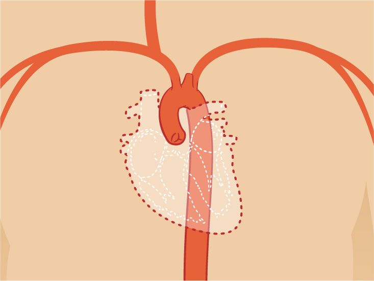
The aorta is the largest artery in the body. It receives oxygen-rich blood from the left ventricle and sends it into smaller arteries that supply the brain, organs, muscles, and tissues. In many heart diagrams, the aorta appears as a large curved vessel at the top of the heart.
Coronary Arteries: The Heart’s Own Blood Supply
The heart pumps blood for the whole body, but the heart muscle also needs its own oxygen supply. That job belongs to the coronary arteries. These vessels wrap around the outside of the heart and deliver oxygen-rich blood to the heart muscle, also called the myocardium.
The two main coronary arteries are the right coronary artery and the left coronary artery. They branch into smaller vessels that supply different areas of the heart. Coronary artery diagrams are especially important because they help explain coronary artery disease, which happens when plaque builds up in the arteries that supply the heart muscle.
When a diagram shows coronary arteries, it may look like the heart is wearing a network of tiny roads. Those roads are not decorative. They are essential. If a coronary artery becomes narrowed or blocked, part of the heart muscle may not receive enough oxygen-rich blood.
The Heart’s Electrical System
The heart is not just a pump; it is also an electrical masterpiece. Its rhythm is controlled by electrical signals that coordinate when the atria and ventricles contract. A heart electrical-system diagram often labels the sinoatrial node, atrioventricular node, bundle branches, and Purkinje fibers.
The sinoatrial node, often called the SA node, is the heart’s natural pacemaker. It starts the electrical signal that causes the atria to contract. The signal then moves to the atrioventricular node, or AV node, and travels through pathways that help the ventricles contract. This carefully timed sequence helps blood move efficiently from chamber to chamber and out to the lungs or body.
How to Read a Heart Diagram Without Panic
Heart diagrams can look overwhelming at first, especially when they include arrows, labels, colors, valves, chambers, and vessels all packed into one image. The trick is to read the diagram in layers.
Step 1: Find the Right and Left Sides
In many medical diagrams, the heart’s right side appears on the left side of the image because you are looking at the heart as if it belongs to someone facing you. This can feel backward at first. Check the labels instead of trusting your first impression.
Step 2: Locate the Four Chambers
Find the right atrium, right ventricle, left atrium, and left ventricle. Remember that atria are on top and ventricles are below. The left ventricle usually has the thickest muscular wall because it pumps blood to the whole body.
Step 3: Follow the Arrows
Arrows show blood-flow direction. Start with blood entering the right atrium from the vena cava. Follow it to the right ventricle, lungs, left atrium, left ventricle, and aorta. If you can trace that path, you understand the main story of circulation.
Step 4: Identify the Valves
Look for the four valves and notice where they sit. The tricuspid and mitral valves are between atria and ventricles. The pulmonary and aortic valves are located where blood leaves the ventricles.
Common Heart Picture Types
Different heart pictures serve different purposes. A classroom diagram may simplify anatomy to help beginners. A medical illustration may add detail for patients or healthcare students. An echocardiogram image shows the heart in motion using ultrasound. A CT or MRI image may show detailed structures from different angles.
For general education, labeled diagrams are usually best. For understanding disease, a comparison picture can be useful. For example, an image comparing a normal valve with a narrowed or leaky valve can make heart valve disease easier to understand. For learning blood flow, arrows and color coding are extremely helpful.
Normal Heart Anatomy vs. Heart Conditions
Understanding normal heart anatomy makes it easier to understand common heart problems. Heart valve disease, for example, may involve valves that do not open fully or do not close tightly. Coronary artery disease involves the arteries that supply blood to the heart muscle. Heart rhythm problems involve the electrical system. Heart failure means the heart is not pumping as effectively as the body needs.
A diagram is not a diagnosis, but it can help explain what a diagnosis means. If a doctor says “mitral valve regurgitation,” a picture can show that the mitral valve is between the left atrium and left ventricle. If someone mentions the aorta, a diagram shows why this large vessel is so important. Visual learning turns medical vocabulary from alphabet soup into something much more digestible.
Quick Anatomy Guide: What to Look for in a Heart Picture
- Chambers: Right atrium, right ventricle, left atrium, left ventricle.
- Valves: Tricuspid, pulmonary, mitral, and aortic valves.
- Major vessels: Vena cava, pulmonary arteries, pulmonary veins, and aorta.
- Septum: Wall separating the right and left sides of the heart.
- Coronary arteries: Vessels that supply the heart muscle.
- Electrical system: Structures that coordinate heartbeat rhythm.
Experience-Based Learning: Making Heart Pictures Actually Useful
Many people first meet heart anatomy through a textbook diagram, and the first reaction is usually not “Ah yes, elegant cardiovascular engineering.” It is more like, “Why does this look like a subway map designed by a very intense doctor?” That reaction is normal. Heart anatomy becomes easier when you connect the picture to real-life experiences.
One practical experience is comparing several heart diagrams instead of relying on one. A simple school diagram might show blue blood on the right side and red blood on the left side. A more detailed medical picture may include the coronary arteries, valves, and electrical pathways. Looking at both helps you understand the difference between a beginner-friendly illustration and a more realistic anatomy image. The simple diagram gives you the map; the detailed one gives you the neighborhood.
Another helpful experience is tracing blood flow with your finger. Start at the vena cava, move into the right atrium, pass through the tricuspid valve, enter the right ventricle, go through the pulmonary valve, travel to the lungs, return through the pulmonary veins, enter the left atrium, pass through the mitral valve, enter the left ventricle, and exit through the aorta. Doing this several times turns memorization into movement. Your brain likes movement. It says, “Finally, something more exciting than staring at labels.”
People also learn heart anatomy better when they connect diagrams to familiar body experiences. For example, feeling your pulse at your wrist connects to the left ventricle pushing blood into the arteries. Hearing a heartbeat during a checkup connects to valves closing during the pumping cycle. Feeling your heart beat faster after running connects to the electrical system and the body’s need for more oxygen-rich blood. These everyday experiences make the diagram feel less like a flat picture and more like a living process.
Patients often find diagrams useful during medical appointments. When a clinician explains a valve problem, a coronary artery blockage, or an abnormal rhythm, a visual can reduce confusion. Instead of trying to imagine invisible structures, the patient can point to the diagram and ask, “Is this the valve you mean?” or “Is this where the artery narrows?” Good diagrams support better questions, and better questions often lead to better understanding.
For students, the best strategy is to redraw the heart from memory. It does not need to be art-gallery quality. A potato-shaped heart with four labeled chambers and arrows can still teach the concept. The goal is not to draw a perfect heart. The goal is to understand how the parts work together. Add the valves, major vessels, and blood-flow arrows. Then compare your sketch with a reliable heart diagram. Every correction helps the anatomy stick.
For writers and educators, heart pictures should be chosen carefully. A beautiful image is not automatically a useful one. The best educational heart diagram has clear labels, accurate blood-flow direction, readable colors, and enough detail for the audience. Beginners need clarity more than complexity. Advanced learners may need coronary arteries, conduction pathways, or cross-sectional views. Matching the picture to the reader is half the job.
The heart is a remarkable organ, but it becomes much easier to understand when pictures, diagrams, and plain-language anatomy work together. Once you know the chambers, valves, vessels, coronary arteries, and electrical system, the heart is no longer a mystery hidden in the chest. It is a powerful, organized pump with excellent timing and surprisingly good teamwork. Not bad for something that never gets a coffee break.
Conclusion
Heart pictures, diagrams, and anatomy guides help explain one of the body’s most important organs in a way that is visual, practical, and memorable. By learning the four chambers, four valves, major blood vessels, coronary arteries, and electrical system, you can understand how the heart moves blood to the lungs and body. A clear heart diagram is more than a picture. It is a roadmap to circulation, health education, and smarter conversations about the cardiovascular system.