
Kidney stones are tiny mineral deposits with a wildly inflated sense of importance. Some remain quietly inside the kidneys, bothering nobody. Others travel into the urinary tract and produce pain so intense that even people with impressive vocabularies may temporarily communicate using only groans.
Although kidney stones can be frightening, they are usually treatable. Small stones may pass with conservative care, while larger, infected, or obstructing stones may require medication or a procedure. Understanding what causes kidney stones, how symptoms develop, and when to seek urgent treatment can make the experience safer and slightly less mysterious.
What Are Kidney Stones?
Kidney stones, medically known as renal calculi or nephrolithiasis, are hard deposits made from minerals, salts, and other substances found in urine. They may form in one or both kidneys and range from microscopic crystals to masses large enough to block urine flow.
The kidneys continuously filter the blood, remove waste, balance minerals, and produce urine. Normally, stone-forming substances remain dissolved. Trouble begins when urine contains more of these substances than its available liquid can comfortably handle. Crystals form, stick together, and gradually build a stonethe urinary system’s least welcome craft project.
Why Kidney Stones Hurt
A stone sitting quietly inside the kidney may cause no symptoms. Pain often begins when it moves into a ureter, one of the narrow tubes carrying urine from the kidneys to the bladder. The ureter may contract around the stone, while urine backs up above the blockage and stretches the kidney’s drainage system.
This combination produces renal colic: severe pain that commonly arrives in waves. The pain may begin below the ribs in the back or side and then move toward the lower abdomen, groin, or genital area as the stone travels.
The Four Main Types of Kidney Stones
1. Calcium Stones
Calcium stones are the most common category. Most contain calcium oxalate, although some are composed mainly of calcium phosphate. Oxalate is naturally present in the body and in foods such as spinach, nuts, beets, chocolate, and certain beans.
Despite the name, people with calcium oxalate stones should not automatically remove calcium-rich foods from their diets. Adequate dietary calcium can bind oxalate in the intestines, reducing the amount that reaches the urine. A severely low-calcium diet may actually increase calcium oxalate stone risk.
2. Uric Acid Stones
Uric acid stones tend to form when urine is persistently acidic. Risk may increase with gout, metabolic conditions, dehydration, or diets containing large amounts of high-purine animal foods, including organ meats, red meat, and certain seafood.
Unlike many other stones, some uric acid stones can be dissolved by raising urine pH with prescribed alkalinizing treatment. This should be medically supervised rather than attempted through an enthusiastic but scientifically questionable home-remedy marathon.
3. Struvite Stones
Struvite stones are associated with urinary tract infections caused by certain bacteria. They may grow rapidly and can become large, branching stones that occupy substantial portions of the kidney. Treatment generally involves controlling the infection and removing as much stone material as possible.
4. Cystine Stones
Cystine stones are uncommon and result from cystinuria, an inherited disorder that causes excessive cystine to enter the urine. Because the underlying condition is lifelong, affected people may develop recurrent stones and often need specialized hydration, diet, medication, and monitoring plans.
What Causes Kidney Stones?
There is rarely one single cause. Kidney stones usually develop through a combination of concentrated urine, individual body chemistry, diet, medical conditions, medications, genetics, and environmental factors.
Not Drinking Enough Fluid
Low urine volume is one of the most important modifiable risk factors. When the body is dehydrated, urine becomes more concentrated, allowing minerals to meet, mingle, and form crystals. Risk may increase during hot weather, strenuous exercise, outdoor work, vomiting, diarrhea, or any routine that makes regular drinking inconvenient.
A High-Sodium Diet
Consuming too much sodium can increase the amount of calcium excreted in urine. Packaged meals, restaurant foods, processed meats, canned soups, salty snacks, and sauces are common sources. The saltshaker may look innocent, but processed food often performs most of the sodium mischief before dinner reaches the table.
Dietary Patterns
High intake of animal protein may raise the risk of uric acid stones and contribute to urine changes that favor certain calcium stones. Large amounts of added sugar may also be unhelpful. However, prevention is not simply a matter of banning every food containing calcium, oxalate, or purines. The right diet depends on the stone’s composition and the patient’s urine chemistry.
Medical Conditions and Digestive Problems
Obesity, diabetes, gout, metabolic syndrome, overactive parathyroid glands, chronic diarrhea, inflammatory bowel disease, and some forms of kidney disease can increase stone risk. Gastric bypass and other intestinal surgeries may change the absorption of calcium, oxalate, fluid, and other substances, making stones more likely.
Family History and Previous Stones
Kidney stones can run in families. A personal history is also important because people who have formed one stone are more likely to form another. Recurrent stones deserve a prevention plan based on evidence rather than a vague instruction to “drink more water and hope for the best.”
Medications and Supplements
Certain medications and supplements can promote stones by changing urine chemistry or crystallizing directly in the urinary tract. Examples may include some diuretics, antivirals, antibiotics, seizure medications, calcium-based antacids, and high-dose vitamin C supplements. Patients should not stop prescribed medicine without consulting the prescriber.
Common Kidney Stone Symptoms
Symptoms depend on the stone’s size, location, movement, and whether infection or obstruction is present. A stone that remains in the kidney may be discovered accidentally during imaging for an unrelated problem.
- Sharp pain in the back, side, lower abdomen, or groin
- Pain that rises and falls in waves
- Pink, red, or brown urine caused by blood
- Burning or pain during urination
- A frequent or urgent need to urinate
- Passing only small amounts of urine
- Cloudy or unusually bad-smelling urine
- Nausea or vomiting
- Restlessness or difficulty finding a comfortable position
Kidney stone pain can resemble appendicitis, gallbladder disease, muscle strain, ovarian conditions, testicular problems, or urinary infection. Severe flank or abdominal pain should therefore be evaluated rather than diagnosed through an online symptom checklist alone.
Warning Signs That Need Urgent Care
Seek immediate medical attention for fever or chills with suspected kidney stone symptoms, inability to urinate, severe or uncontrolled pain, repeated vomiting, faintness, confusion, or significant weakness. Urgent evaluation is also important for people who are pregnant, have one functioning kidney, have reduced kidney function, or are immunocompromised.
A blocked urinary system combined with infection is an emergency. Antibiotics alone may not be enough if infected urine cannot drain. Doctors may need to place a ureteral stent or a drainage tube through the back before treating the stone itself.
How Kidney Stones Are Diagnosed
Medical History and Physical Examination
A clinician will ask where the pain began, whether it moves, how suddenly it started, and whether it is accompanied by urinary symptoms, fever, nausea, or vomiting. Previous stones, family history, dietary habits, medical conditions, medications, and supplements can provide useful clues.
Urine and Blood Tests
Urinalysis may detect blood, infection, crystals, urine acidity, and other abnormalities. Blood tests can evaluate kidney function and measure substances such as calcium and uric acid. People with recurrent or high-risk stones may be asked to complete one or more 24-hour urine collections.
Imaging Tests
Imaging can reveal a stone’s location, size, number, and effect on urine drainage. A noncontrast CT scan provides detailed information and is frequently used in adults with acute symptoms. Ultrasound avoids ionizing radiation and may be preferred in situations such as pregnancy or pediatric evaluation. The choice depends on the patient and clinical circumstances.
Stone Analysis
A patient may be asked to urinate through a strainer and save any stone that passes. Laboratory analysis identifies its composition and helps guide prevention. One person may need to reduce sodium, another may need urine alkalinization, and a third may need treatment for an infection or inherited disorder.
Kidney Stone Treatment Options
Treatment depends on the stone’s size, location, composition, associated symptoms, likelihood of passing, and whether it is causing infection, bleeding, obstruction, or kidney dysfunction.
Conservative Treatment for Small Stones
A small stone may pass naturally if pain is controlled, urine can flow, and there is no evidence of infection or serious kidney impairment. A clinician may recommend appropriate pain medication, regular fluid intake, anti-nausea medicine, and follow-up imaging.
Some patients with ureteral stones receive an alpha blocker to relax the ureter and improve the chance of passage. This treatment is not right for every stone or every patient, so it should be prescribed after medical assessment.
Extracorporeal Shock Wave Lithotripsy
Extracorporeal shock wave lithotripsy, commonly shortened to ESWL or SWL, directs energy waves toward a stone from outside the body. The waves break the stone into smaller fragments that can pass in urine. Success depends on factors such as stone size, hardness, location, anatomy, and patient health.
Ureteroscopy
During ureteroscopy, a urologist passes a thin scope through the urethra and bladder into the ureter or kidney. The stone may be removed with a small basket or fragmented with laser energy. A temporary ureteral stent may be placed to maintain drainage while swelling resolves.
Percutaneous Nephrolithotomy
Percutaneous nephrolithotomy, or PCNL, is commonly used for very large, complex, or difficult-to-reach kidney stones. The surgeon enters the kidney through a small incision in the back and removes the stone using specialized instruments. It is more invasive than ureteroscopy or shock wave treatment but can be the most effective approach for a substantial stone burden.
Medication Based on Stone Type
Preventive or stone-specific medications may include potassium citrate to raise urinary citrate or pH, thiazide-type medicine for certain patients with excessive urinary calcium, or allopurinol for selected people with recurrent stones related to uric acid production. Infection stones require appropriate antimicrobial care, while cystine stones may need specialized medications.
How to Help Prevent Kidney Stones
Produce Plenty of Urine
Prevention guidelines commonly focus on drinking enough fluid to produce approximately 2 to 2.5 liters of urine daily. The exact amount a person must drink varies with climate, exercise, body size, diet, pregnancy, illness, and medical restrictions. People with heart failure, advanced kidney disease, or fluid limits should ask their clinicians for individualized advice.
Keep Normal Calcium-Rich Foods
Unless a clinician recommends otherwise, calcium should generally come from a balanced diet rather than being eliminated. Eating calcium-containing food with meals can help trap dietary oxalate in the digestive tract before it reaches the kidneys. Calcium supplements require individualized advice because dose, timing, and medical history matter.
Reduce Excess Sodium
Read nutrition labels and compare serving sizes. Choosing fresh foods more often, rinsing canned products, limiting processed meats, and using herbs or acids for flavor can lower sodium intake without turning dinner into a plate of nutritional sadness.
Do Not Declare War on Every Oxalate Food
A strict low-oxalate diet is not automatically necessary. Many oxalate-containing foods also provide fiber, magnesium, potassium, and other nutrients. People with high urinary oxalate may be advised to limit especially concentrated sources while eating adequate calcium with meals.
Moderate Animal Protein and Added Sugar
Depending on the stone type, reducing oversized portions of red meat, organ meat, processed meat, and certain seafood may help. Eating more fruits, vegetables, legumes, and whole grains can support a urine environment that is less welcoming to many stones.
Request a Personalized Evaluation After Recurrent Stones
People with repeated stones, unusual stone types, a family history, a single kidney, intestinal disease, or stones beginning at a young age may benefit from specialist evaluation. Stone analysis, blood testing, and 24-hour urine studies can identify specific targets for diet or medication.
Frequently Asked Questions
Can kidney stones go away without treatment?
Some small stones pass without a procedure. Others become lodged, cause persistent pain, block urine, or contribute to infection. Size is important, but location, anatomy, symptoms, and medical history also influence the chance of passage.
Does drinking water dissolve kidney stones?
Water helps prevent stones by diluting urine and may support the natural passage of a small stone, but it does not dissolve most existing calcium stones. Some uric acid stones can be dissolved by medically raising urine pH.
How long does passing a kidney stone take?
The timeline varies from days to several weeks. A stone’s size and location strongly affect passage, but there is no reliable universal countdown. Follow-up is important because a decrease in pain does not always prove that the stone has left the urinary tract.
Are kidney stones the same as gallstones?
No. Kidney stones form in the urinary system, while gallstones form in the gallbladder and involve bile. Both may cause dramatic abdominal pain, but they have different causes, locations, and treatments.
A Realistic Kidney Stone Experience: From First Pain to Prevention
The following is a composite example based on common kidney stone experiences. It is not the story of a specific patient, and individual symptoms and treatment decisions vary.
The First Twinge
Imagine waking with a dull ache beneath the ribs on one side. At first, it feels like a muscle strainperhaps the reward for sleeping in a position normally attempted only by folding chairs. Stretching does not help. Twenty minutes later, the ache becomes a sharp, gripping pain that rises, fades, and returns.
The person cannot get comfortable. Sitting feels wrong. Standing feels wrong. Lying down feels like an administrative error. Soon, the pain begins moving toward the lower abdomen, and nausea joins the party without being invited.
Deciding Whether to Seek Care
A first-time episode of severe flank pain deserves medical evaluation because many conditions can produce similar symptoms. In this example, the person notices pink urine and vomits after trying to drink water. A family member drives them to urgent care or an emergency department rather than allowing someone in severe pain to drive.
At the clinic, the staff checks temperature, blood pressure, pain level, and medical history. A urine sample shows blood but no obvious sign of infection. Blood tests assess kidney function, and imaging reveals a small stone in the ureter with mild backup of urine.
The Treatment Conversation
Because the stone appears likely to pass, the clinician recommends conservative management. The patient receives appropriate pain relief, anti-nausea treatment, instructions about fluid intake, and possibly a prescription to relax the ureter. They are also given a urine strainer and clear warning signs that require an immediate return.
This conversation matters. “You have a kidney stone” is not a complete plan. Patients should understand where the stone is, approximately how large it is, whether urine flow is obstructed, whether infection is suspected, what medicines to take, and when follow-up should occur.
The Waiting Period
At home, discomfort comes in waves. The patient takes medication only as directed and drinks regularly rather than attempting to swallow a swimming pool in one sitting. They monitor their temperature and urine output and use the strainer each time they urinate.
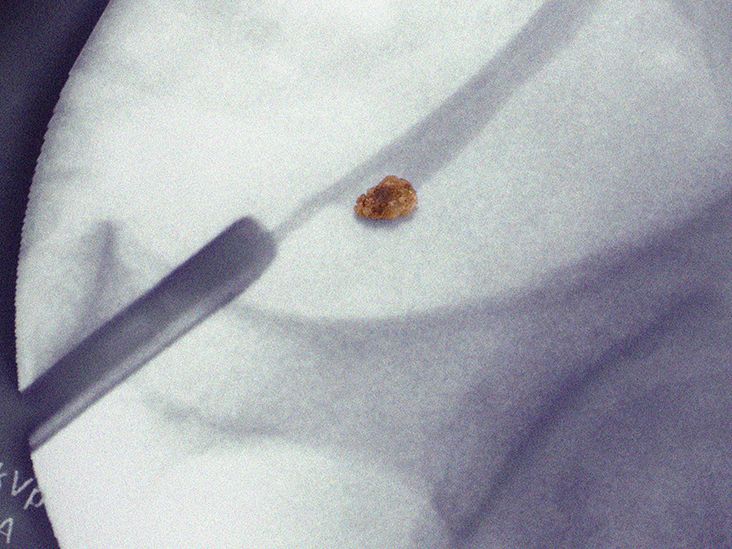
Two days later, the pain shifts lower. The patient feels urinary urgency and then spots a tiny, rough object in the strainer. It looks almost offensively small compared with the trouble it caused. The stone is placed in a clean container for laboratory analysis.
What Happens After the Stone Passes
Passing the stone is relief, not necessarily the end of care. The clinician may arrange follow-up imaging to confirm that obstruction has resolved. Laboratory analysis identifies a calcium oxalate stone. Because this was a first episode without unusual risk factors, the initial prevention plan focuses on fluid intake, moderate sodium, normal dietary calcium, balanced protein portions, and more fruits and vegetables.
If stones recur, the evaluation becomes more detailed. A 24-hour urine collection may reveal low urine volume, high urinary calcium, low citrate, excessive oxalate, abnormal acidity, or another treatable pattern. The plan can then be adjusted instead of relying on a generic forbidden-food list downloaded from the internet.
Lessons From the Experience
The most useful lesson is not simply that kidney stones hurt. It is that symptoms should be taken seriously, infection must be ruled out, and prevention works best when it is personalized. Keeping a refillable water bottle nearby, checking urine color, reducing sodium, and following through with testing may be much less exciting than emergency pain treatmentand that is precisely the point.
Conclusion
Kidney stones form when minerals and other substances crystallize in concentrated urine. Dehydration, excessive sodium, certain dietary patterns, genetics, infections, digestive disorders, metabolic conditions, medications, and inherited diseases can all contribute.
Severe side or back pain, blood in the urine, nausea, urinary urgency, and painful urination are common symptoms. Fever, chills, inability to urinate, persistent vomiting, or uncontrolled pain require urgent medical attention.
Treatment ranges from pain control and observation to shock wave lithotripsy, ureteroscopy, or percutaneous surgery. After the immediate crisis, stone analysis and targeted prevention can reduce the chance of another unwelcome mineral reunion. Hydration matters, but the smartest long-term plan is one matched to the stone type, urine chemistry, medical history, and nutritional needs.