
Acral lentiginous melanoma can develop on the palms, soles, or beneath the nailsplaces that rarely headline anyone’s skin-check routine. Knowing its subtle warning signs can lead to earlier diagnosis, more treatment options, and a better outlook.
What Is Acral Lentiginous Melanoma?
Acral lentiginous melanoma, commonly abbreviated as ALM, is a rare subtype of melanoma that begins in melanocytes, the pigment-producing cells of the skin. “Acral” refers to the hands, feet, fingers, toes, and nails, while “lentiginous” describes the pattern in which abnormal melanocytes initially spread along the skin’s surface.
Unlike most cutaneous melanomas, acral lentiginous melanoma usually develops in areas that receive little ultraviolet exposure. Its typical locations include:
- The soles of the feet, particularly weight-bearing areas and the heels
- The palms of the hands
- Beneath a fingernail or toenail, where it may be called subungual melanoma
- The skin around a nail
ALM can affect people of every skin color. Although melanoma overall is diagnosed more frequently in White Americans, acral melanoma accounts for a larger proportion of melanoma cases among Black, Asian, and Hispanic populations. This distinction matters: darker skin does not provide immunity from melanoma, especially on the palms, soles, and nails.
The disease is often discovered later than other melanomas because it may resemble a bruise, wart, callus, fungal infection, or minor injury. Unfortunately, suspicious spots do not carry tiny signs saying, “Hello, I require a biopsy.” Awareness therefore plays an important role in early detection.
What Causes Acral Lentiginous Melanoma?
The precise cause is not fully understood. Regular ultraviolet exposure is a major risk factor for many melanomas, but it does not appear to be the primary driver of ALM. A person can practice excellent sun safety and still develop this cancer.
Researchers have identified genetic changes within some acral melanoma tumors. Compared with common sun-related melanomas, ALM is less likely to carry a BRAF mutation and may be more likely to contain alterations involving genes such as KIT or structural changes involving larger sections of DNA. These findings help explain why molecular testing may influence treatment choices in advanced disease.
Pressure, friction, and injuries have been investigated as possible contributors, particularly because many tumors appear on weight-bearing portions of the foot. However, ordinary trauma has not been proven to cause acral lentiginous melanoma. An injury may simply draw attention to a lesion that was already developing.
Who Is at Risk?
ALM is more commonly diagnosed in middle-aged and older adults, although it can occur at almost any age. It affects all racial and ethnic groups. Traditional melanoma risk factorssuch as fair skin, frequent sunburns, and extensive UV exposureare not reliable predictors for this particular subtype.
The most important practical lesson is simple: everyone should include palms, soles, toes, fingers, and nails in routine skin checks. Melanoma apparently did not receive the memo that it was supposed to stay on sun-exposed skin.
Acral Lentiginous Melanoma Symptoms
The first symptom is usually a new or changing area of pigmentation. The lesion may be brown, black, gray, blue, red, or a mixture of colors. Some acral melanomas contain little pigment and appear pink, red, or skin-colored, making them easier to overlook.
Signs on the Soles or Palms
A suspicious lesion on a palm or sole may have one or more of the following characteristics:
- A new dark patch that slowly becomes wider
- An irregular shape or uneven border
- Several shades within the same spot
- A mark that looks noticeably different from nearby spots
- A pink, red, or colorless growth that enlarges
- A sore that does not heal normally
- Bleeding, cracking, itching, tenderness, or ulceration
- A mass or “bruise” that persists after an injury should have healed
The familiar ABCDE warning signsasymmetry, border irregularity, color variation, diameter, and evolutioncan be helpful. Evolution is especially important: any spot that changes in size, shape, color, texture, or symptoms deserves attention. However, ALM does not always follow the standard ABCDE script, so a small or evenly colored lesion should not automatically receive a free pass.
Symptoms Under a Nail
Subungual melanoma often begins as a dark vertical streak running from the base toward the tip of a nail. Warning signs may include:
- A new brown or black band, especially in one nail
- A band that becomes wider, darker, or more irregular
- Pigment that is widest near the cuticle
- Color spreading from the nail onto the cuticle or surrounding skin
- A nail that cracks, splits, lifts, or becomes distorted
- A lump, sore, or area of bleeding beneath the nail
- Persistent pain without a clear explanation
Pigment extending onto the surrounding nail skin is known as Hutchinson sign. It can raise concern for melanoma, but it is not present in every case and can occasionally occur in benign conditions. Likewise, many dark nail streaks are harmless. The appearance alone cannot confirm the diagnosis.
When Should You See a Dermatologist?
Arrange an evaluation if a new lesion persists, grows, bleeds, changes, or fails to heal. A spot initially blamed on a blister, bruise, splinter, or fungus should be rechecked if it does not improve as expected. New or changing nail bands also deserve assessment, particularly when only one nail is affected.
Do not cut, burn, freeze, or repeatedly treat an unexplained lesion with over-the-counter products before obtaining a diagnosis. Home surgery is rarely a good hobby, and it is especially unhelpful when melanoma is a possibility.
How Acral Lentiginous Melanoma Is Diagnosed
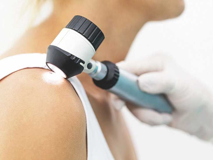
A dermatologist begins by examining the lesion and the rest of the skin, including all nails. A dermoscopea handheld magnifying instrument with specialized lightingcan reveal pigment patterns that are difficult to see with the unaided eye.
Photography may document the lesion’s size and appearance, but photographs and phone applications cannot rule out cancer. If melanoma is suspected, a biopsy is necessary.
Skin and Nail Biopsy
During a biopsy, a clinician removes tissue for examination by a dermatopathologist. When feasible, the entire suspicious lesion is removed with a narrow margin. Larger lesions or those in anatomically difficult locations may require a carefully selected partial biopsy that includes the most concerning area.
Nail-unit biopsies require particular expertise because the tumor may begin in the nail matrix, where the nail grows. Sampling the wrong location can produce an incomplete answer. For a suspicious nail lesion, referral to a dermatologist or surgeon experienced with nail disorders can be valuable.
Understanding the Pathology Report
If melanoma is confirmed, the pathology report may describe:
- Breslow thickness: How deeply the melanoma has grown into the skin
- Ulceration: Whether the surface over the tumor has broken down
- Mitotic activity: Evidence of how actively cells are dividing
- Margins: Whether melanoma cells reach the edges of the removed tissue
- In situ or invasive disease: Whether abnormal cells remain in the epidermis or have entered deeper tissue
Breslow thickness and ulceration are especially important for staging and prognosis.
Tests for Possible Spread
Doctors may discuss a sentinel lymph node biopsy for an invasive melanoma based on its thickness, ulceration, and other risk features. This procedure removes the first lymph node or nodes to which cancer would be most likely to travel.
Imaging tests such as CT, PET, or MRI scans are generally reserved for higher-stage disease, concerning symptoms, or findings suggesting spread. Early, thin melanomas usually do not require a grand tour of every scanner in the hospital.
Stages of Acral Lentiginous Melanoma
ALM uses the same general staging system as other cutaneous melanomas:
- Stage 0: Melanoma in situ, limited to the outermost skin layer
- Stages I and II: Invasive melanoma confined to the original area, with stage determined mainly by thickness and ulceration
- Stage III: Cancer has reached nearby lymph nodes, lymphatic channels, or nearby skin
- Stage IV: Cancer has spread to distant organs or distant areas of skin or lymph nodes
Stage is more informative than the label “acral” alone when doctors plan treatment and discuss an individual outlook.
Acral Lentiginous Melanoma Treatment
Treatment depends on the stage, tumor location, molecular features, general health, and effects that surgery may have on hand or foot function. Care may involve dermatology, surgical oncology, medical oncology, plastic surgery, podiatry, rehabilitation, and other specialties.
Surgery for Localized Disease
Wide local excision is the main treatment for localized ALM. The surgeon removes the melanoma along with a margin of normal-appearing tissue. The required margin depends largely on tumor thickness.
Because palms, soles, and nail units are functionally important areas, reconstruction may require a skin graft or tissue flap. Amputation is not automatically required for nail melanoma. When possible, specialists use tissue-sparing approaches that completely remove the cancer while preserving function.
Immunotherapy
People with high-risk, unresectable, recurrent, or metastatic melanoma may receive immune checkpoint inhibitors. Medicines such as pembrolizumab or nivolumab help immune cells recognize and attack cancer. Nivolumab may also be combined with ipilimumab in selected cases, although combination treatment can cause more serious immune-related side effects.
Immunotherapy may be used after surgery to reduce recurrence risk, before surgery in certain advanced but operable cases, or as treatment for metastatic disease. ALM can respond to immunotherapy, although response patterns may differ from those seen in heavily sun-damaged melanoma.
Targeted and Other Treatments
Tumor testing can identify actionable mutations. A BRAF V600 mutation may make BRAF and MEK inhibitor combinations an option. Some acral melanomas have KIT alterations, but not every alteration responds to a KIT-directed medicine. The exact molecular finding matters.
Radiation therapy may help control a difficult local tumor, lymph-node disease, or symptoms from metastases. Surgery, clinical trials, and newer treatments may also be considered. Lifileucel, a tumor-infiltrating lymphocyte therapy, is an option for certain adults with unresectable or metastatic melanoma that has progressed after specified prior treatments.
Acral Lentiginous Melanoma Prognosis
ALM is highly treatable and often curable when found early. Its overall statistics have historically been less favorable than those of more common melanoma subtypes, largely because acral tumors are frequently diagnosed after they have become thicker or spread. Delayed recognition, access to dermatologic care, tumor biology, and health disparities can all contribute.
Factors associated with prognosis include:
- Stage at diagnosis
- Breslow thickness
- Presence or absence of ulceration
- Lymph-node involvement
- Whether the cancer has reached distant organs
- Whether surgery can completely remove the tumor
- The tumor’s molecular characteristics and response to treatment
- The person’s overall health
Current American Cancer Society figures for melanoma overall report five-year relative survival of more than 99% for localized disease, approximately 76% for regional disease, and about 35% for distant disease. These are population estimates for all cutaneous melanoma typesnot ALM-specific predictionsand cannot determine what will happen to one person.
Treatments for advanced melanoma have improved substantially, so older statistics may not fully reflect outcomes for people receiving modern immunotherapy, targeted therapy, or cellular therapy. A clinician familiar with the pathology, stage, scans, and treatment response can provide a much more meaningful prognosis.
Can Acral Lentiginous Melanoma Be Prevented?
Because ALM is not clearly driven by sun exposure, sunscreen cannot specifically guarantee its prevention. Sun protection remains important for preventing other UV-related skin cancers, however, and should remain part of a sensible skin-care routine.
Early detection is the most practical defense. Once a month, examine:
- Both palms and the spaces between the fingers
- Fingernails, toenails, and surrounding skin
- The tops, sides, heels, and soles of both feet
- The spaces between the toes
Use a mirror or ask a trusted person for help with hard-to-see areas. Photographing a questionable spot beside a ruler can make genuine changes easier to identify. If something evolves, show the images to a dermatologist rather than conducting a six-month internet debate with yourself.
Experiences Related to Acral Lentiginous Melanoma
The experience of ALM often begins with uncertainty rather than an obvious cancer warning. The following examples are composites based on commonly reported diagnostic and treatment experiences. They do not describe specific individuals, but they illustrate why persistence and specialist evaluation matter.
The “Bruise” That Does Not Leave
Imagine noticing a dark mark on your heel after buying new shoes. A bruise seems logical, so you wait. Several weeks later, the mark remains and appears slightly wider. It does not hurt, which feels reassuringbut melanoma can be painless. A primary care clinician suspects friction and recommends observation. When the area is still present two months later, a dermatologist examines it with a dermoscope and recommends a biopsy.
Many patients describe this stage as emotionally confusing. The lesion looks ordinary enough to dismiss, yet the word “biopsy” immediately turns the mental volume to maximum. The useful lesson is not that every bruise is cancer. It is that a presumed bruise should behave like one: it should gradually fade or move outward as a nail grows. A persistent or evolving mark deserves another look.
Waiting for the Pathology Results
The biopsy itself is often less difficult than anticipated. Local anesthetic can sting briefly, and a foot biopsy may require temporary activity adjustments or special wound care. The waiting period is frequently harder. Patients may inspect an online portal every few minutes, despite knowing that pathology reports do not mature faster when stared at.
If melanoma is diagnosed, unfamiliar terms can make the report sound like a foreign language. Asking the clinician to explain Breslow thickness, ulceration, margins, and stage in plain English can restore a sense of control. Writing down questions or bringing another person to appointments helps, especially when anxiety makes details difficult to retain.
Balancing Cancer Control and Function
Treatment on a thumb, toe, palm, or sole raises practical concerns. Patients often ask whether they will be able to walk comfortably, work with their hands, exercise, drive, or return to a physically demanding job. These questions are not cosmetic side notes; they are central to quality of life.
A multidisciplinary team can plan cancer removal and reconstruction together. Depending on the tumor, recovery may involve dressings, limited weight-bearing, a graft, physical therapy, occupational therapy, or footwear modifications. Healing can feel frustratingly slow because hands and feet are involved in nearly everything. Following wound-care instructions and reporting infection, worsening pain, or graft changes promptly can prevent avoidable setbacks.
Living After Treatment
Finishing treatment does not always produce instant emotional relief. Some survivors feel anxious whenever they notice a new spot, bruise, or nail streak. Follow-up appointments can create “scanxiety,” even when previous examinations have been reassuring.
A structured plan often helps. Patients can ask who will perform skin examinations, how often lymph nodes or scans will be checked, which symptoms require an urgent call, and whether family members need routine dermatologic care. Monthly self-exams are useful, but checking several times each day usually feeds anxiety without improving detection.
Support groups, oncology social workers, counselors, rehabilitation professionals, and trusted family members can make recovery less isolating. The goal is not to become a full-time detective assigned to every freckle. It is to build a calm, repeatable routine that catches meaningful changes while allowing ordinary life to resume.
Questions to Ask Your Healthcare Team
- Is the melanoma in situ or invasive?
- What are its Breslow thickness, ulceration status, and stage?
- Do I need a sentinel lymph node biopsy?
- How will surgery affect walking, hand function, or nail growth?
- Should my tumor undergo molecular testing?
- Could I benefit from immunotherapy, targeted therapy, or a clinical trial?
- What follow-up schedule and self-examination routine do you recommend?
Conclusion
Acral lentiginous melanoma is uncommon, but its hiding places are familiar: palms, soles, and nails. A changing dark patch, unexplained growth, persistent sore, or evolving nail streak should be examined rather than endlessly treated as a bruise or fungus.
A biopsy is the only reliable way to diagnose ALM. When the disease is found while localized, surgery can often be curative. Advanced cases may require immunotherapy, targeted treatment, radiation, cellular therapy, or participation in a clinical trial. Prognosis depends most strongly on tumor depth and stage, making timely evaluation the most valuable step a person can take.