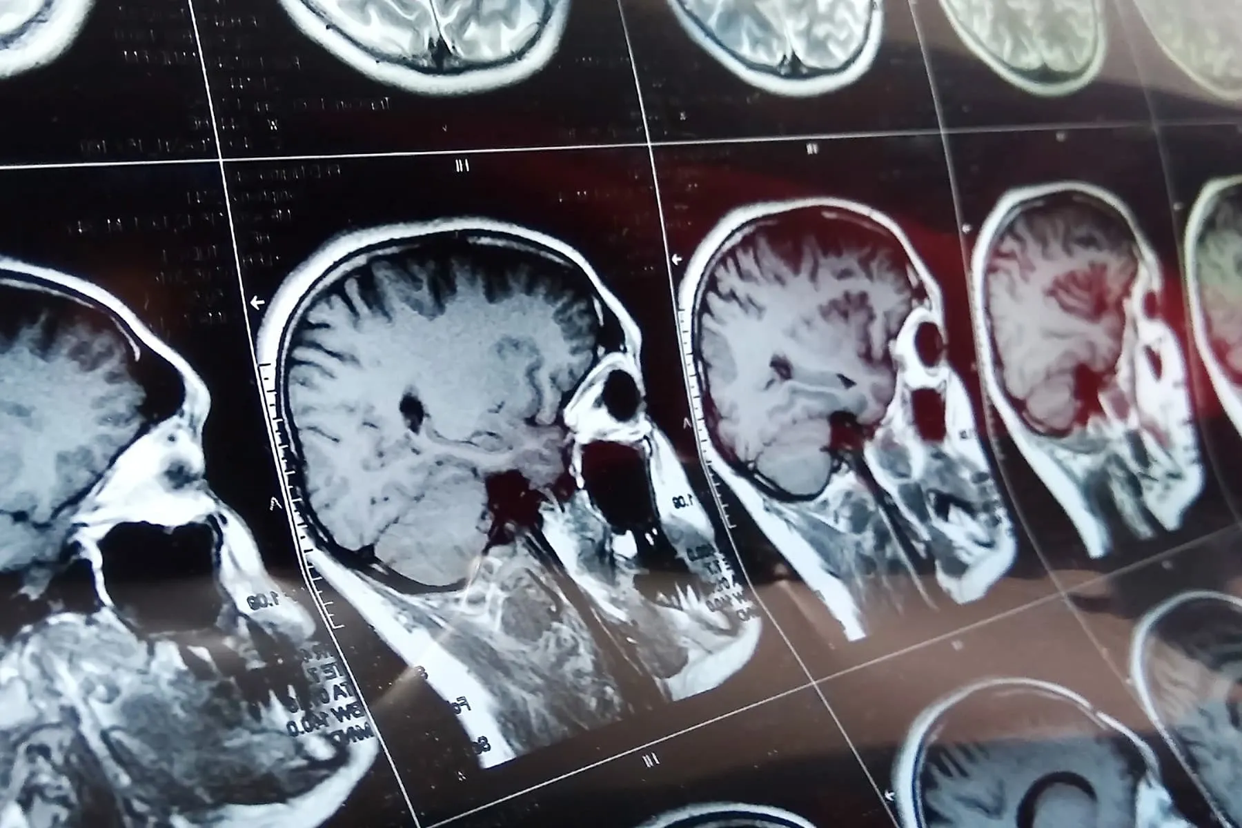

It sounds like something from a medical drama that got slightly carried away in the writers’ room: doctors performing brain surgery on a baby who had not even been born yet. But this was not a cliffhanger, a sci-fi subplot, or a heroic surgeon shouting “scalpel” under dramatic lighting. It was a real medical milestone involving a rare and dangerous condition called vein of Galen malformation, and it gave one baby a chance at life before she ever took her first breath.
The headline “Doctors Perform First Surgery on Baby’s Brain in the Womb” refers to a groundbreaking fetal procedure performed by teams from Boston Children’s Hospital and Brigham and Women’s Hospital. The patient was Denver Coleman, a baby diagnosed before birth with a severe vascular abnormality in her brain. Instead of waiting until deliverywhen babies with this condition can crash quickly from heart failure or brain injurydoctors intervened while she was still in the womb.
The procedure was not “brain surgery” in the old-fashioned open-skull sense. No tiny fetal operating room. No miniature surgeon’s cap. Instead, it was an extremely precise, ultrasound-guided fetal embolization. Doctors inserted a needle through the uterus, reached the abnormal blood vessel area in the fetal brain, and placed tiny coils to reduce dangerous blood flow. The goal was simple but ambitious: prevent the newborn emergency before it happened.
What Is Vein of Galen Malformation?
Vein of Galen malformation, often shortened to VOGM, is a rare congenital blood vessel abnormality that develops during fetal life. In a typical brain, arteries carry oxygen-rich blood into smaller vessels called capillaries, which slow the flow and allow oxygen to nourish tissue. In VOGM, some arteries connect directly to a large deep vein instead of passing through those capillaries.
That direct connection creates a high-pressure, high-flow situationbasically a plumbing problem in the most delicate plumbing system imaginable. Blood rushes too fast into veins that were never designed to handle arterial pressure. The baby’s heart may work overtime trying to keep up, and the brain may be exposed to abnormal pressure and circulation.
For newborns with severe VOGM, the first hours or days after birth can be extremely dangerous. Once the placenta is no longer helping manage circulation, the baby’s heart may struggle under the burden. Complications can include high-output heart failure, pulmonary hypertension, brain injury, seizures, hydrocephalus, developmental delays, and death.
Why the Womb Made a Difference
Traditionally, doctors treated vein of Galen malformation after birth, often with embolization procedures that block or reduce abnormal blood flow. In many cases, postnatal treatment can work well. But severe fetal cases are different. Some babies are already showing signs of strain before birth, and the transition from womb to world can be brutal.
Inside the womb, the placenta acts like a powerful support system. It helps manage the baby’s circulation and can buffer some of the abnormal blood-flow effects. After birth, that backup system is gone. For a baby with a severe VOGM, the heart may suddenly face a workload it cannot tolerate.
This is why the Boston team’s approach was so important. The logic was not merely “operate earlier.” It was “change the circulation before the crisis begins.” By reducing the abnormal flow before delivery, doctors hoped to give the baby’s heart and brain a calmer start. In medical terms, that is elegant. In parent terms, it is the difference between entering the world with a fighting chance and entering it already in emergency mode.
The Case of Baby Denver Coleman
Denver Coleman’s pregnancy had been progressing normally until a routine ultrasound late in pregnancy showed something unusual. Further imaging revealed a severe vein of Galen malformation. Her parents, Kenyatta and Derek Coleman, were suddenly facing a terrifying decision: wait until birth and hope postnatal treatment could happen fast enough, or enroll in a first-of-its-kind clinical trial.
The procedure took place at about 34 weeks of pregnancy. Using ultrasound guidance, the medical team carefully navigated a needle through the mother’s abdomen and uterus and into the fetus’s skull. Through that access, doctors placed tiny metal coils into the abnormal vessel area to slow the excessive blood flow.
The results were remarkable. Imaging showed a rapid reduction in abnormal flow, and fetal heart measurements improved. Denver was born shortly afterward and did not require the kind of immediate emergency heart-failure treatment doctors often fear in severe VOGM cases. She spent time in the neonatal intensive care unit for monitoring, but early reports described her as eating, gaining weight, and progressing well.
How Fetal Embolization Works
Fetal embolization is the technical heart of this breakthrough. Embolization means placing material inside a blood vessel to reduce or block abnormal blood flow. In adults and children, embolization is commonly performed through catheters inserted into blood vessels. In a fetus, the challenge becomes far more complex because doctors must safely access a tiny moving patient inside another patient.
During Denver’s procedure, specialists used ultrasound as their navigation system. The mother received care from maternal-fetal medicine experts, while pediatric anesthesiology helped keep the fetus still and comfortable. Neurointerventional specialists then delivered coils into the malformation to reduce the dangerous rush of blood.
The goal was not to completely shut down every abnormal vessel in one dramatic move. That could create new risks. Instead, doctors aimed to reduce the flow enough to protect the heart and brain during birth and the newborn period. Think of it less like turning off a faucet and more like carefully lowering a fire hose from “blast through a wall” to “manageable stream.”
Why This Was a Medical First
This surgery was widely described as the first successful in-utero brain procedure of its kind for vein of Galen malformation. It represented a new frontier in fetal therapy, a field that already includes prenatal treatment for conditions such as spina bifida, twin-to-twin transfusion syndrome, and certain fetal airway or heart problems.
What made this case different was the target: a blood vessel malformation deep inside the fetal brain. That required collaboration across multiple specialtiesmaternal-fetal medicine, pediatric neurosurgery, neurointerventional radiology, fetal imaging, anesthesiology, neonatology, cardiology, and nursing. One doctor alone could not pull this off. This was less “lone genius with a laser pointer” and more “medical orchestra trying to play Beethoven during an earthquake.”
The procedure also changed the conversation around VOGM. Instead of seeing fetal diagnosis only as a warning sign before postnatal treatment, doctors began exploring whether prenatal intervention could prevent some of the worst damage from happening in the first place.
What Later Research Has Shown
The first case was not the end of the story. Early trial data later reported outcomes from a small group of fetuses with severe vein of Galen malformation. In that early experience, several fetal embolizations were completed successfully, and surviving treated babies showed encouraging neurological development in early follow-up.
However, the results also came with important caution. This is still an emerging therapy. Some babies in the early trial did not survive, and unscheduled or preterm delivery was a major concern. That matters because fetal surgery always involves two patients: the pregnant person and the fetus. Any benefit must be weighed against risks such as preterm birth, bleeding, infection, fetal distress, anesthesia complications, and the emotional strain of high-stakes decision-making.
In other words, this is not a simple miracle button. It is a promising, highly specialized procedure for carefully selected cases. Families should not read about it and assume every VOGM diagnosis should be treated before birth. The right plan depends on imaging, fetal heart function, brain findings, gestational age, hospital expertise, and whether the condition appears severe enough to justify intervention.
How Doctors Diagnose VOGM Before Birth
Many cases of vein of Galen malformation are first suspected during prenatal ultrasound. Doctors may see an abnormal fluid-filled structure in the fetal brain or signs that the baby’s heart is enlarged or working too hard. Doppler ultrasound can help detect unusual blood flow patterns.
Fetal MRI may then provide more detail about the brain, blood vessels, and whether there is already evidence of injury. Fetal echocardiography can assess the baby’s heart function. Together, these tools help doctors decide whether the baby is stable enough to wait for treatment after delivery or at high risk of rapid decline after birth.
That level of imaging is crucial because VOGM exists on a spectrum. Some babies have less severe forms and can be treated after birth with good outcomes. Others have aggressive, high-flow malformations that place the heart and brain at serious risk even before delivery.
The Emotional Side of a Prenatal Brain Diagnosis
For parents, hearing that an unborn baby has a rare brain condition is devastating. Pregnancy is already full of strange math, snack cravings, and late-night questions like “Is this normal?” Add a diagnosis involving the fetal brain and heart, and suddenly the entire future feels like it has been placed under hospital lighting.
Parents may have to make decisions quickly, often while learning unfamiliar medical language. Terms like “embolization,” “fetal MRI,” “high-output heart failure,” and “neurodevelopmental outcome” do not exactly roll gently into family conversations. A good care team must do more than offer technical skill. They must translate, support, repeat information, and recognize that families are trying to make life-changing decisions while scared.
This case showed the power of clear communication. Denver’s parents were not choosing between an easy option and a hard option. They were choosing between unknowns. That is often the reality of cutting-edge medicine: hope arrives wearing a lab coat, but uncertainty comes in the same room.
Why This Breakthrough Matters Beyond One Baby
Denver’s story matters because it suggests a possible shift in how doctors think about certain fetal brain conditions. The old model was: diagnose before birth, prepare for emergency treatment after birth. The new question is: can some emergencies be softened or prevented before birth?
If fetal embolization continues to show safety and benefit in larger studies, it could become an option for the most severe VOGM cases. It may also encourage researchers to explore other prenatal vascular interventions. Medicine often advances this way: one careful case, one small trial, one question that refuses to stay theoretical.
Still, responsible optimism is important. A breakthrough is not the same as a cure. A first success does not erase risk. The most honest takeaway is that fetal brain surgery for VOGM has moved from “almost unimaginable” to “possible in expert hands for selected patients.” That is a huge leap, even if the road ahead remains long.
What Families Should Know About This Procedure
It is not available everywhere
This kind of fetal brain procedure requires an elite multidisciplinary center with experience in fetal surgery, neurointerventional radiology, pediatric neurology, fetal imaging, and neonatal intensive care. Most hospitals cannot offer it, and that is not a criticism. Most hospitals also do not keep a spaceship in the basement.
It is designed for severe cases
Not every baby with VOGM is a candidate. Some babies may do better with careful monitoring and treatment after delivery. Doctors must evaluate the malformation’s size, flow, heart effects, fetal brain findings, and overall pregnancy status.
It carries real risks
Potential benefits include reducing heart failure and brain injury after birth. Potential risks include preterm delivery, fetal complications, procedure failure, and maternal complications. The best decisions come from detailed conversations with specialists who understand both fetal therapy and VOGM.
Experiences and Reflections Related to Doctors Performing Brain Surgery on a Baby in the Womb
When people read a headline like “Doctors Perform First Surgery on Baby’s Brain in the Womb,” the first reaction is usually a stunned pause. Then comes the question: how is that even possible? That reaction is completely fair. Most of us think of surgery as something that happens in an operating room to a patient lying on a table. Fetal surgery rewrites that picture. The patient is hidden, tiny, developing, and protected inside another person’s body. The surgeon cannot simply “look directly” at the target. Every movement must be guided by imaging, planning, and teamwork.
For families who have experienced complicated pregnancies, this story may feel both inspiring and emotionally intense. A routine ultrasound can change everything in minutes. One moment, parents are wondering about nursery colors or whether the baby will inherit Dad’s eyebrows. The next, they are hearing about brain blood flow, fetal heart strain, and survival odds. That emotional whiplash is difficult to describe unless you have lived through a serious prenatal diagnosis.
Doctors and nurses who work in fetal care often talk about the importance of balancing hope with honesty. Families need hope because without it, decision-making becomes unbearable. But they also need honesty because medical risk cannot be softened into a fairy tale. In Denver Coleman’s case, the outcome was widely described as extraordinary, but it was extraordinary precisely because the condition was so dangerous.
There is also an experience here for the medical community. Breakthroughs like this do not appear overnight. They come from years of anatomy research, imaging improvements, fetal therapy experience, device development, ethical review, trial planning, and careful patient selection. Before the public sees the dramatic headline, many scientists and clinicians have spent years asking boring-looking questions that save lives later. Medical progress is often less fireworks and more spreadsheets, simulations, meetings, and someone saying, “Let’s check that measurement again.”
For readers, the most meaningful lesson may be that prenatal medicine is becoming more proactive. Not long ago, many fetal diagnoses mainly helped doctors prepare for birth. Now, in select cases, doctors may be able to treat a condition before birth changes the baby’s physiology for the worse. That does not mean every condition can or should be treated in the womb. But it does mean the womb is no longer viewed only as a waiting room. In advanced fetal centers, it can sometimes become the first place healing begins.
Parents facing a VOGM diagnosis should take away neither panic nor unrealistic expectation. Instead, they should take away one practical message: seek evaluation at a center with deep expertise. Ask about fetal MRI, fetal echocardiography, postnatal embolization, delivery planning, clinical trials, and long-term neurological follow-up. The best care plan is rarely built from one dramatic procedure. It is built from a team that knows what to do before birth, during delivery, after birth, and throughout childhood.
Conclusion
The first successful surgery on a baby’s brain in the womb was more than a headline built for clicks. It was a turning point in fetal medicine. By treating a severe vein of Galen malformation before birth, doctors showed that it may be possible to reduce life-threatening blood-flow problems before a newborn’s heart and brain are overwhelmed.
The story of Denver Coleman is hopeful, but it is not simple. Fetal brain embolization remains a specialized, emerging therapy with real risks and unanswered questions. Early research is promising, especially for the most severe cases, but larger studies and long-term follow-up are essential.
Still, the achievement deserves awe. A baby’s brain was treated before birth. A family was given hope in a situation that once offered very little. And medicine took one careful step into a future where some of the most dangerous newborn emergencies may be addressed before the first cry.
Note: This article is for informational and educational purposes only. It is based on real public medical reporting and research about fetal embolization for vein of Galen malformation. It should not replace medical advice from qualified maternal-fetal medicine, pediatric neurosurgery, cardiology, or neonatal specialists.